OBSERVATIONS ON THE pH OF THE ARTERIAL BLOOD, THE pH AND THE ELECTRICAL ACTIVITY OF THE CEREBRAL CORTEX12 [26]
J.G. Dusser de Barenne, Clyde S. Marshall, W.S. McCulloch and Leslie F. Nims
##
In this paper will be presented some results of experiments in which the pH of the arterial blood, the pH of the cerebral cortex and the electrical activity of the cortex were recorded simultaneously under various experimental conditions.
The techniques employed have been fully described in previous papers. Specially constructed glass electrodes in conjunction with vacuum tube-microvoltmeters allow continuous photographic records of the pH of the blood and the cortex. The electrical activity of the cortex was recorded separately with Ag-AgCl2 electrodes through AC-amplifiers and a Westinghouse four-element oscillograph.
With these techniques it can be shown (I) that under certain conditions changes in the pH of the cortex are parallel to those in the arterial blood; (II) that under other conditions changes in the pH of the cortex are not parallel to those of the arterial blood.
I. A striking instance of the first group of conditions can be obtained in experiments in which the artificial respiration is changed in the curarized animal. In such experiments a decrease of the artificial respiration promptly results in an acid shift in the pH of the blood and cortex. After restoration of the artificial respiration to the initial volume the pH of both blood and cortex rapidly returns to its original level. The changes in the pH of the cortex lag slightly behind those of the blood and are usually less rapid. Obviously under these conditions the pH of the cortex passively follows that of the arterial blood. See Figure 1.
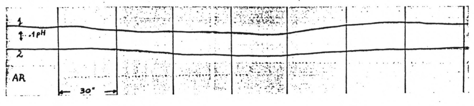
Figure 1. Curarized dog under artificial respiration (AR). Record 1 = pH of arterial blood (femoral artery). Record 2 - pH of cerebral cortex. Note that both pH of blood and cortex shift towards acid side upon decrease of artificial respiration and return towards previous pH level upon restoration of original volume of the artificial respiration, blood made non-coagulable by intravenous injection of chlorazol fast pink.
II. That the pH of the cortex need not follow the pH of the arterial blood can be inferred from observations in which the pH of one portion of the cortex is changed by local treatment which leaves the pH of the rest of the cortex unchanged. Local thermocoagulation (1, 2) or local electrical after-discharge following electrical stimulation (2) gives a local acid shift in the area involved; local freezing or local application of iodoacetic acid gives a local alkaline shift in the area involved. See Figure 2.
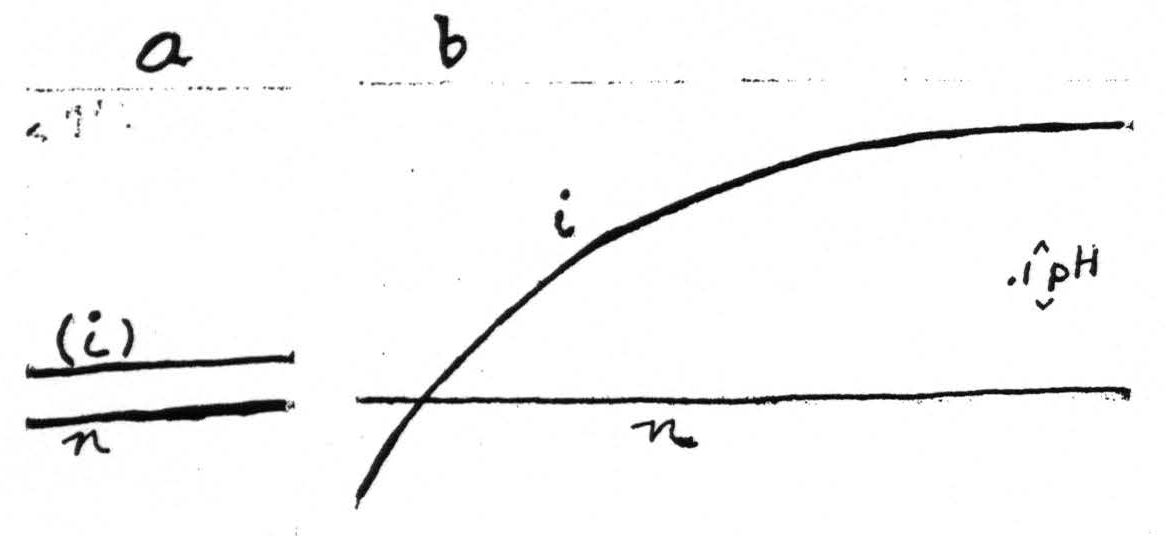
Figure 2. Shows the local development of alkalinity (record i) in an area of the cerebral cortex (monkey) treated with local application of iodoacetic acid (between sections a and b). pH of an adjacent untreated area remains unchanged (record n).
Direct evidence that the pH of the cortex does not always parallel that of the arterial blood can be obtained in experiments with intravenous injection of monobromide of camphor.
Figure 3 shows the pH of the arterial blood and of the cortex and the simultaneous electrical activity of the cortex in a curarized dog under constant artificial respiration before and after an intravenous injection of camphor sufficiently large to produce a “central” epileptoid discharge of the cortex. It will be seen, (1) that under these conditions the records of the pH of the cortex and of the blood do not parallel each other; (2) that the record of the pH of the blood presents a steady slow acid shift, whereas the pH of the cortex, ca 80 seconds after the injection, shows a marked alkaline wave followed by a larger enduring acid wave and a subsequent return towards its original level at a time when the blood is still “going acid”.
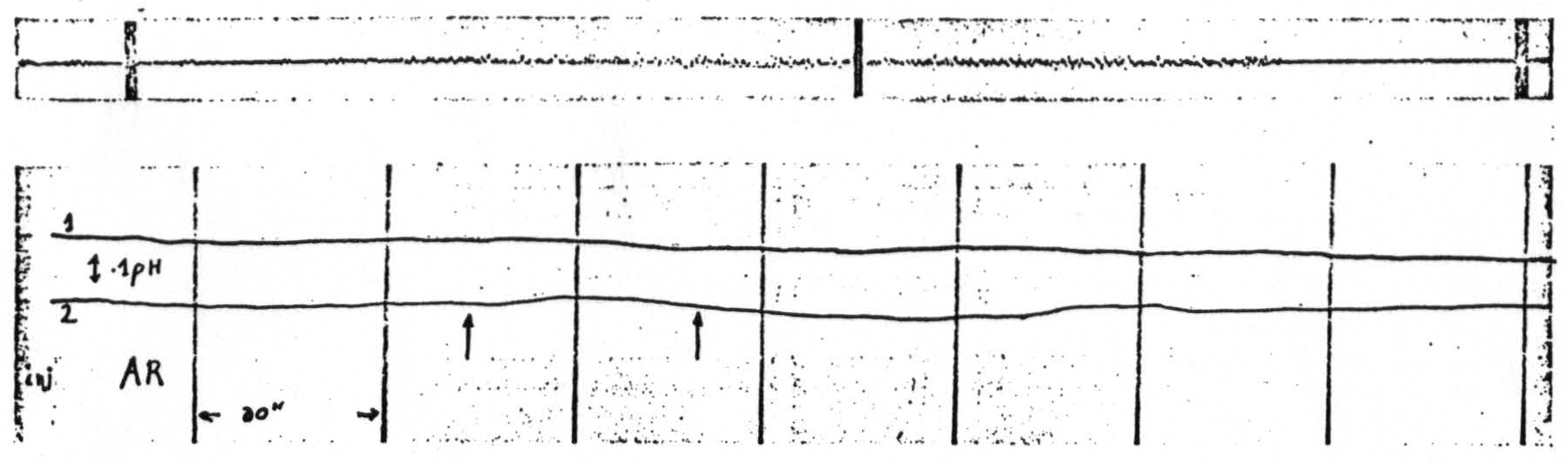
Figure 3. The upper record shows an epileptoid electrical discharge in a (sensory) area of the cortex of a curarized dog following an intravenous injection of monobromide of camphor. The lower record shows the changes in pH of the blood (1) and of an area of the cortex (2) as close as convenient to that mentioned above. Note the alkaline wave followed by an acid swing in the record of the cortical pH and the non-parallelism of the two pH-curves. The period of the seizure is marked in the lower record by the two arrows. Time-markings in the two records coincide (30 sec.). AR = record of artificial respiration. Blood rendered non-coagulable by intravenous injection of chlorazol fast pink.
Figure 4 shows the pH records of the pre- and postcentral arm-areas and the electrical activity of these areas in a curarized monkey under constant artificial respiration following an intravenous injection of monobromide of camphor, which resulted in a “central” epileptoid discharge. It should be noted, (1) that the changes in the two pH records although of the same direction do
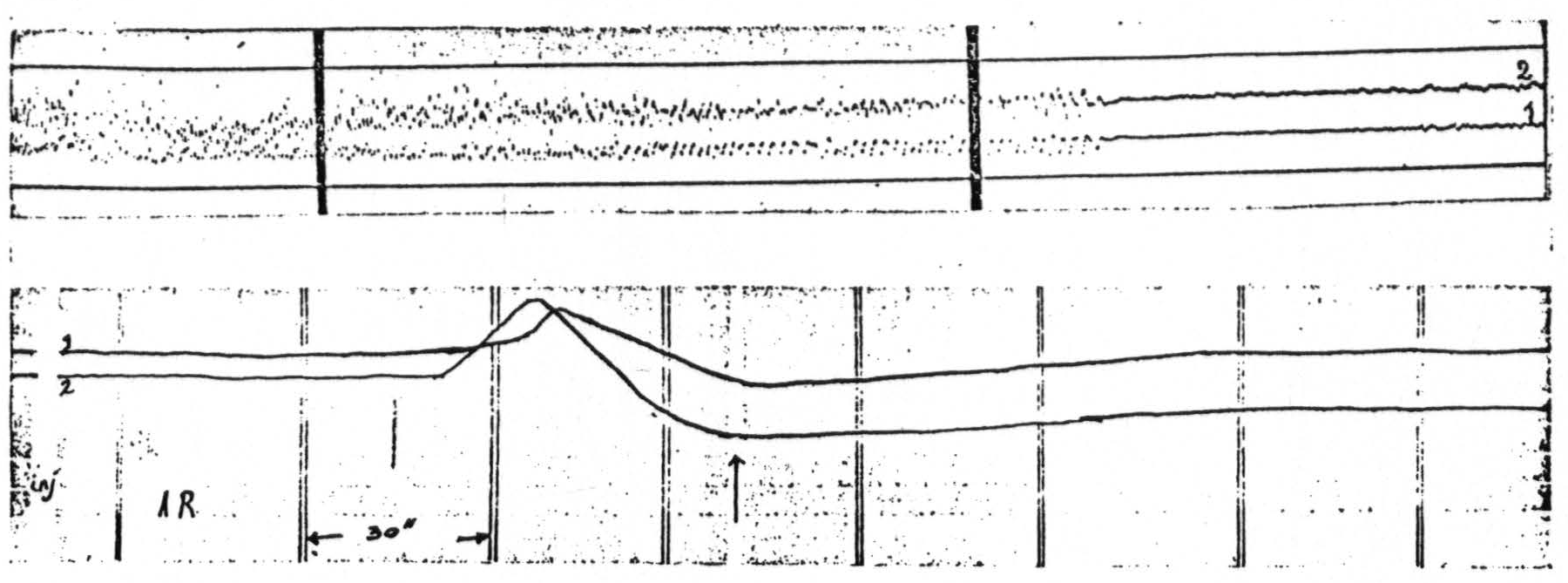
Figure 4. The upper section shows the almost complete course (except for the very beginning) of a “central” epileptoid attack in a curarized monkey as reflected in the electrocorticograms of a pre- and postcentral area of the arm region, elicited by an intravenous injection of 1.2 mgm. of monobromide of camphor. 1 = record of electrocorticogram of A.4, 2 = record of electrocorticogram of A.1-2. The lower section of the figure shows the changes in pH of these same areas before, during and after the seizure. This began ca 55 seconds after the intravenous injection. The period of the seizure is indicated in the lower section of the figure by the two arrows. The time lines in the two records coincide (30 sec.). AR in lower section is record of the artificial respiration.
not parallel each other exactly; (2) that the changes are much greater than those in Figure 3, probably due to the fact that the epileptoid cortical discharge was much more severe in the experiment on the monkey.
From these records it seems permissible to conclude that the initial alkaline shift of the pH of the cortex is associated with the onset of the “central” epileptoid seizure and as the seizure proceeds the cortex becomes “acid”.
Taken together these observations show that as soon as any alteration in the activity of the cortex occurs, its pH no longer passively reflects that of the arterial blood.
Footnotes
References:
Dusser de Barenne, J.G., W.S. McCulloch and LF. Nims. Proc. Soc. Experi. Biol. Med. 36:462,1937.
Dusser de Barenne, J.G., W.S. McCulloch and LF. Nims. J. Cell. and Comp. Physiol. 10:277, 1937.
Nims, LF. Yale J. Biol. Med. 10:241, 1938.
Nims, LF., C. Marshall and H.S. Burr. Science 87: 197, 1938.
Nims, LF. and C. Marshall. Yale J. Biol. Med. 10:445, 1938.
Marshall, C. and LF. Nims. Yale J. Biol. Med.10:561, 1938.
For further research:
Wordcloud: Acid, Activity, Alkaline, Area, Arterial, Artificial, Barenne, Biol, Blood, Camphor, Central, Cerebral, Changes, Conditions, Cortex, Curarized, Discharge, Dog, Dusser, Electrical, Epileptoid, Experiments, Figure, Following, Injection, Intravenous, Local, Lower, Marshall, McCulloch, Med, Monkey, Monobromide, Nims, Observations, Parallel, Ph, Record, Respiration, Results, Return, Section, Seizure, Shift, Shows, Towards, Wave, Yale
Keywords: Cortex, Blood, Changes, Experiments, Conditions, Tube-Microvoltmeters, Vision, Electrodes, Knowledge
Google Books: http://asclinks.live/qyzu
Google Scholar: http://asclinks.live/6awb
Jstor: http://asclinks.live/zqmr